
What is a Mole Check?
A mole check is a medical examination of the skin by a dermatologist to assess moles and other skin lesions for signs of skin cancer, particularly melanoma.
Moles, also known as nevi, are common skin growths that usually appear as small, dark brown spots. Moles form when melanocytes, the cells that produce pigment, grow in clusters or clumps. Sun exposure and genetic factors can influence the number and appearance of moles. Most people have between 10 and 40 moles, which can develop anywhere on the skin, including the scalp, armpits, under the nails, and between fingers and toes.
Moles, although mostly harmless, are important indicators. Normal moles will be small, with even, well-defined borders. They are usually tan, light brown, or medium brown in color, and generally less than a quarter of an inch in size. Abnormal moles are larger, with a diameter of over 6 millimeters. They will have ragged, poorly defined borders, and they may itch, bleed, or ooze fluid. If you have concerns about a specific mole or notice changes, it is important to seek advice from a healthcare professional, preferably a dermatologist.
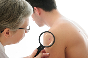
What Happens During Mole Checks?
During a mole check, a dermatologist or trained healthcare professional will:
- Medical History Review: Ask about your medical history, including any personal or family history of skin cancer, your history of sun exposure, and any changes you have noticed in your moles.
- Visual Inspection: Examine the skin for any abnormal moles or lesions. This includes noting their size, shape, color, and any changes over time.
- Dermatoscopy: Use a dermatoscope, a specialized magnifying tool, to get a closer look at the moles. This tool allows the examiner to see patterns and structures in the skin that are not visible to the naked eye.
- ABCDE Rule: Evaluate moles using the ABCDE rule:
- Asymmetry: One half of the mole does not match the other half.
- Border: Edges are irregular, ragged, or blurred.
- Color: Color is not uniform and may include different shades of brown, black, pink, red, white, or blue.
- Diameter: The mole is larger than 6 millimeters (about the size of a pencil eraser).
- Evolving: The mole changes in size, shape, color, or begins to bleed or itch.
- Palpation: Feel the moles to check for any lumps or changes in texture.
- Photographs: Take photos of the moles to track changes over time.
After the examination, the dermatologist will discuss their findings with you, provide any necessary recommendations for treatment or follow-up, and offer advice on skincare and sun protection. If any moles appear suspicious, the dermatologist may recommend a biopsy, where a small sample of the mole is removed and examined under a microscope to check for cancerous cells. Regular mole checks are important for early detection of skin cancer, especially for individuals with a high number of moles, a history of sun exposure, or a family history of skin cancer.
For more information about our services or to schedule an appointment, click here to request an appointment online. We’ll respond to you as soon as possible.